
O-scan SMART
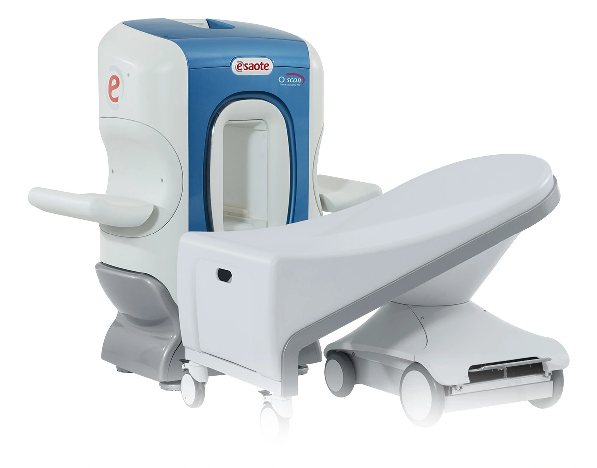
The O-scan SMART from Esaote is an extremity MRI system designed for dedicated MSK imaging, simplified workflow, and patient comfort.
O-scan SMARTTechnical Specifications
| System Type | Extremity MRI system |
|---|---|
| Display | Real-time monitor for direct positioning checks from the gantry |
| Imaging Technology | Dedicated extremity MRI; real-time positioning; optional eXP technology with SpeedUp, TR reduction, 3D imaging, True-Motion, and metal artifact reduction |
| Image Processing | AgilExam optional slice auto-alignment for automatic positioning by laterality and anatomy size |
| Workflow Interface | Real-time positioning monitor; AgilExam three-click workflow for exam task selection and slice confirmation |
| Software Platform | Optional eXP technology platform; optional AgilExam workflow package |
| Dimensions / Size | Fits in 9 m² / 100 ft²; with RF cage: 3 m × 4 m; without RF cage: 2.8 m × 3.7 m |
| Power / Charging | Plug-and-play installation; low power consumption; helium-free MRI system |
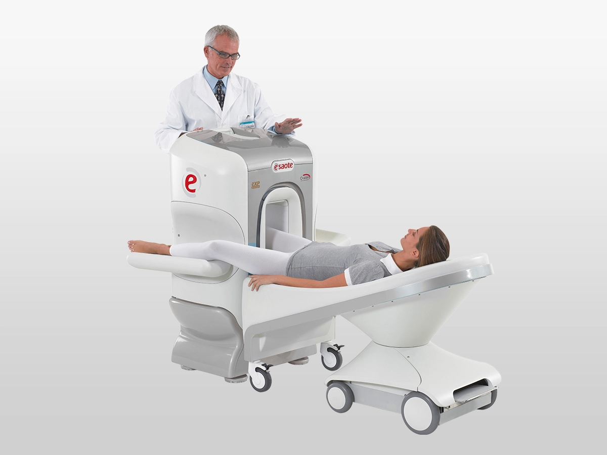
| Mounting / Configuration | Available with or without RF cage; only the extremity under investigation is placed in the system |
| Compatibility / Retrofit | Can operate as a standalone specialty MRI or alongside whole-body scanners in radiology departments |
| Imaging Capability | Dedicated imaging for joints and limbs; 3D SHARC cartilage imaging; 3D isotropic 0.4 mm; X-Bone; metal artifact reduction; True-Motion dynamic joint imaging |
| Clinical Applications | Knee, ankle, foot, wrist, hand, elbow, calf, sports medicine, podiatry, rheumatology, and post-surgical extremity MRI |
Dedicated MSK MRI Efficiency
O-scan SMART combines dedicated extremity MRI, compact installation, and streamlined MSK workflow.
Compact Extremity Workflow
O-scan SMART is built for small-footprint installation and plug-and-play operation. Real-time positioning and optional AgilExam help simplify setup and improve throughput.
Advanced Extremity Imaging
The system supports dedicated joint and limb MRI with optional 3D isotropic imaging, cartilage imaging, metal artifact reduction, and dynamic True-Motion assessment.
Designed for Patient Comfort
Only the extremity being examined is placed inside the system, helping reduce claustrophobia. The open, patient-friendly design supports specialty MSK imaging in daily practice.
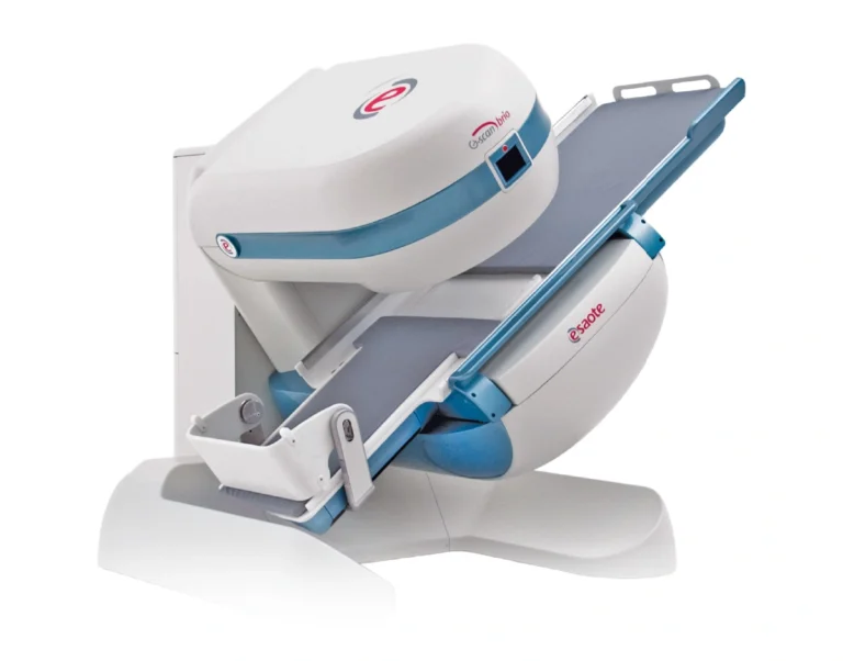
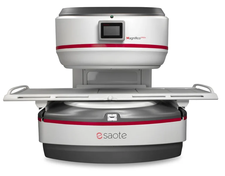
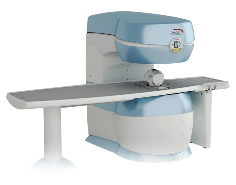
Related Products
Imaging System Support
Your imaging equipment should perform reliably from day one through years of clinical use. Imperial provides ongoing support programs designed specifically for installed systems—helping protect image quality, reduce service disruptions, and extend equipment value.
Installation & System Readiness
Imperial prepares each system for real-world clinical use through coordinated delivery, installation oversight, system testing, and workflow validation to ensure confident day-one operation.
Performance Maintenance
Scheduled service and calibration help maintain imaging accuracy, system stability, and consistent performance across daily patient volumes and evolving clinical demands.
Technical Service Support
When service needs arise, Imperial’s imaging specialists provide dependable technical support focused on restoring performance and minimizing workflow interruption.
Get a Tailored Quote
Tell us about your imaging needs and goals—our team will provide a path forward.
Share your medical imaging equipment, service needs, or growth plans. Imperial will follow up with clear, practical recommendations.