Vet MRI is an increasingly valuable tool in veterinary medicine, providing exceptional soft-tissue detail without the ionizing radiation that makes veterinary radiography safety and CT dose management a concern. But implementing MRI involves more than selecting the right magnet. Patient safety, anesthesia, monitoring, equipment, room design, staff training, and MRI protocols all contribute to a successful veterinary MRI department.
Two safety considerations veterinary teams should understand are radiofrequency (RF) heating and thermal burns.
Because veterinary MRI patients are typically under general anesthesia and cannot communicate discomfort, proper monitoring, positioning, equipment selection, and staff training are especially important.
How Can a Patient Heat Up During an MRI?
MRI uses radiofrequency energy as part of the image-acquisition process. Some of this RF energy is absorbed by the patient's tissues and converted into heat.
MRI systems monitor RF energy deposition using parameters such as Specific Absorption Rate (SAR) or other system-specific RF exposure metrics. The amount of heating can vary depending on the MRI system, field strength, patient size, positioning, coil configuration, and pulse sequences being used.
Veterinary patients present an additional challenge because most dogs and cats undergoing MRI are anesthetized. Anesthesia alters normal thermoregulation, while the patient cannot move or alert the team if something feels unusually warm.
Although hypothermia remains a common concern in anesthetized veterinary patients, MRI teams should understand both sides of temperature management and continuously monitor the patient throughout the procedure.
Ways to Reduce the Risk of Excessive Heating
-
Use MRI-appropriate patient monitoring
Continuous physiologic monitoring is essential during veterinary MRI. Depending on the patient and procedure, this should include heart rate, respiratory rate, oxygen saturation, end-tidal CO₂, blood pressure, and temperature.
Monitoring equipment used within the MRI environment must be appropriate for the magnetic field and configured according to the manufacturer's instructions. As a veterinary equipment supplier, Imperial Imaging recommends MR conditional monitors with compatible pediatric accessories and MR conditional anesthesia machines and ventilators.
-
Select the appropriate coil
Choosing a coil appropriate for the patient's size and anatomy can improve signal-to-noise ratio and may allow the team to obtain diagnostic images more efficiently.
Available coil configurations should be considered when comparing veterinary imaging equipment, not after installation.
-
Optimize veterinary MRI protocols
Not every MRI sequence deposits the same amount of RF energy. Sequence parameters, field strength, scan duration, and other factors can influence RF exposure.
Protocols should therefore be developed specifically for the MRI system, anatomy, patient population, and diagnostic objective.
Experienced MRI applications specialists and veterinary radiologists can help hospitals create protocols that balance:
- Diagnostic image quality
- Scan time
- Anesthesia time
- RF exposure
- Patient safety
- Efficient hospital workflow
-
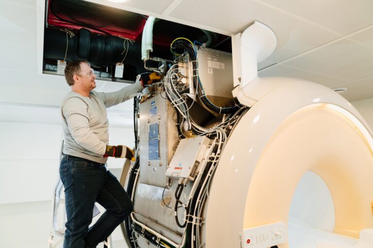
Maintain and service the MRI system
Preventive maintenance should be part of every veterinary MRI program. Hospitals should follow the MRI manufacturer's recommended inspection, testing, and service schedules and address system warnings or abnormalities promptly.
-
Train the entire MRI team
MRI safety should not be limited to the technologist operating the scanner.
Veterinarians, technicians, anesthesia personnel, maintenance staff, and anyone entering controlled MRI areas should understand the hazards associated with the magnetic field and RF environment.
A veterinary hospital should also maintain clearly defined procedures for screening patients, equipment, staff, and visitors before entering the MRI environment.
What Causes MRI Burns?
MRI-related thermal injuries can occur when RF energy produces localized heating.
Metallic or conductive objects are not the only potential cause of burns. Conductive materials or cables, body loops, and improper positioning against equipment or the scanner bore can also cause localized heating.
This is particularly important in veterinary medicine because the patient is anesthetized and unable to communicate discomfort.
Potential hazards can include:
- Metallic foreign bodies
- Certain implants
- ECG leads and cables
- Pulse oximetry equipment
- Conductive monitoring components
- Improperly routed cables
- Some wraps, blankets, or positioning materials
- Patient contact with the MRI bore
- Skin-to-skin contact that creates conductive loops
Even a retained BB or other metallic foreign body may warrant evaluation before entering the MRI environment.
How Can Veterinary Hospitals Reduce MRI Burn Risk?
-
Establish a formal pre-MRI screening process
Every patient should be screened before MRI for implants, previous surgical procedures, metallic foreign bodies, and external equipment.
When the history is uncertain and a metallic foreign body is suspected, additional evaluation, potentially including radiographs, may be appropriate.
-
Pay careful attention to patient positioning
Remove collars, tags, and unnecessary accessories before scanning.
Position and pad the patient according to the MRI manufacturer's safety guidance. Avoid skin-to-skin contact that could create conductive loops and direct contact with the bore when required.
-
Manage monitoring cables carefully
Monitoring cables and leads should be MRI-appropriate and routed according to manufacturer instructions.
Avoid loops, unnecessary cable contact with the patient, and configurations that could contribute to localized heating.
-
Verify every accessory entering the MRI environment
Monitoring equipment, infusion equipment, positioning devices, oxygen components, stretchers, tools, and other accessories entering controlled MRI areas must be evaluated for use in that environment.
Terms such as MR Safe, MR Conditional, and MR Unsafe have specific meanings and should be understood by everyone working around the magnet.
-
Build emergency procedures before the first patient is scanned
Every MRI program should have written procedures addressing emergencies such as:
- Patient instability
- Suspected thermal injury
- Anesthesia complications
- Equipment failure
- Emergency patient removal
- Fire or other facility emergencies
- Magnet-related emergencies
These procedures should be incorporated into staff training rather than developed after an incident occurs.
Veterinary MRI Safety Starts Before Installation
Many MRI safety and workflow problems can be prevented during the planning and equipment-selection stage.
Hospitals comparing veterinary imaging systems should evaluate not only field strength and purchase price, but also:
- Magnet and system configuration
- Available coils
- MRI room and site requirements
- RF shielding
- Electrical and HVAC requirements
- Patient anesthesia and monitoring
- MRI-compatible ancillary equipment
- Patient positioning and workflow
- Applications training
- Veterinary imaging protocols
- Preventive maintenance
- Local and remote service support
- Long-term operating costs
A properly planned MRI suite can improve patient safety while also making studies faster, easier, and more financially sustainable for the hospital.
Building or Upgrading a Veterinary MRI Program?
Imperial Imaging Technology helps veterinary hospitals evaluate and implement veterinary imaging solutions, from new, pre-owned, and refurbished MRI systems to full-body vet digital imaging.
As a full-service veterinary equipment company, our team can assist from equipment selection through site planning, construction coordination, installation, applications training, and ongoing service.
Whether you're purchasing your hospital's first veterinary MRI, replacing an aging system, or considering a refurbished system, choosing the right equipment is only one part of the decision.
The goal is to build an MRI program that produces excellent diagnostic images, protects patients and staff, supports efficient clinical workflow, and makes financial sense for the hospital.
Looking for a Veterinary MRI System?
Talk with Imperial Imaging Technology about veterinary MRI equipment, animal imaging solutions, and support for your imaging project.