In the ever-evolving field of medical technology, radiology stands on the front line of innovation and advancement. From X-ray machines to MRIs and Computer Tomography machines, radiology equipment has created essential solutions for medical diagnostics and treatment planning. So what exactly is radiology and why is it needed? Our seasoned experts at Imperial Imaging Technology would be happy to share their knowledge. Join us below as we discuss the importance of radiology within the healthcare community, how to utilize the most common types of medical imaging systems, and more.
What is Radiology?
Radiology is a medical field that utilizes imaging techniques to diagnose and treat diseases. There are two main types of radiology – diagnostic radiology and interventional radiology. They are distinct but closely related branches of radiology, each playing crucial roles in the diagnosis, treatment, and management of medical conditions.
Diagnostic radiology includes various imaging techniques that allow clinicians to diagnose diseases and conditions within the body. Diagnostic radiology might include traditional X-rays and screening exams such as mammography and Computed Tomography (CT). This field of radiology also allows for treatment planning, as the images obtained provide vital information that guides treatment decisions, surgical planning, and monitoring of treatment responses.
Interventional radiology uses imaging guidance to perform minimally invasive procedures for diagnostic and therapeutic purposes. Radiologists in this field are trained to use techniques such as fluoroscopy and ultrasound to treat targeted areas in the body. Examples of these procedures include biopsy, tumor ablation, and embolization. Interventional radiology fills a gap in the medical field because these treatments provide excellent alternatives to traditional surgery, reducing risks, complications, and recovery times.
Whether diagnostic or interventional, radiology contributes significantly to modern medicine and patient care. So what types of medical imaging systems are used in the field of radiology? Below we will dig deeper into the most common medical imaging systems and what each machine accomplishes.
Radiology & The Use Of Imaging Equipment
Radiology utilizes different types of imaging equipment to visualize internal structures and processes within the body. While each machine maintains different functions, they all contribute to a clinician’s ability to diagnose, plan treatment, monitor diseases, and conduct minimally invasive procedures. Having the necessary high-quality imaging equipment allows healthcare workers to serve patients with excellence.
Magnetic Resonance Imaging (MRI)
Magnetic Resonance Imaging (MRI) machines use strong magnetic fields and radio waves to generate detailed images of the body’s internal structure. This non-invasive imaging technique does not use radiation, making it unique from other devices prominent in radiology – such as the many types of X-ray machines. Clinicians can utilize MRI in a handful of different ways, including:
- Diagnostic Imaging – MRI can evaluate soft tissues and organs such as the brain, spinal cord, and heart. These detailed images can reveal abnormalities such as tumors, inflammation, injuries, and structural anomalies.
- Functional MRI (fMRI) – Functional MRI is a technique that assesses brain activity by detecting changes in blood flow. These results are used in research and clinical settings to study brain functions such as language, movement, and sensory processing.
- Magnetic Resonance Angiography (MRA) – This specialized MRI technique is used to visualize blood vessels and assess blood flow. It is commonly used to diagnose conditions such as aneurysms, stenosis, and vascular malformations.
MRI is essential to radiology because it provides a non-invasive and detailed look at the body’s internal structure. Facilities can offer excellent patient care with the help of advanced MRI machines such as the Esaote G-Scan, Siemens Magnetom Aera, and more.
Ultrasound
Ultrasound machines use high-frequency sound waves transmitted through a probe to create real-time images of organs and tissues. A computer is connected to the probe and uses the sound waves to create the imaging. The common uses of ultrasound include:
- Pregnancy Monitoring – Ultrasounds can be used for monitoring fetal development and detecting abnormalities during pregnancy.
- Abdominal Imaging – Ultrasound machines can also be used to assess organs like the liver, kidneys, and gallbladder for tumors, stones, or fluid collections.
- Vascular Studies – Doppler ultrasound evaluates blood flow in the arteries and veins to identify blockages or narrowing.
Healthcare facilities utilize ultrasound machines because they are safe and non-invasive. They are useful in a wide range of medical fields, spanning from obstetrics and gynecology to cardiology. Among the high-quality ultrasound equipment options on the market are the Mindray Consona N9, Mindray TE7, and more.
X-Ray
X-ray machines emit ionizing radiation to create images of bones, tissues, and organs. They consist of a generator that produces X-rays and a detector (film or digital sensor) to capture the images. This common technology can diagnose fractures, infections, tumors, lung diseases, dental issues, and many other conditions.
There are many types of X-ray machines, including but not limited to:
- U-Arm X-Ray Machine – The U-arm X-ray machine is a sophisticated imaging tool designed with a U-shaped configuration, offering enhanced flexibility and maneuverability in medical imaging procedures. These machines are often used in orthopedic exams, chest X-rays, and extremity imaging.
- Straight Arm X-Ray Machine – The straight arm design of this machine allows for easy positioning of patients and optimal imaging results while also taking up minimal space in your facility. This machine is ideal for Urgent Care, family practices, and beyond.
- C-Arm X-Ray Machine – These versatile machines offer real-time imaging capabilities crucial for surgical interventions, orthopedic procedures, and more. Clinicians can capture high-quality images from multiple angles, enhancing diagnostic accuracy and treatment outcomes.
Mammography
Mammography machines can detect breast cancer by using low-dose X-rays to create detailed images of the breast tissue. The specialized imaging equipment creates images called mammograms that could show precancerous or cancerous conditions. During a mammogram appointment, the breast is compressed between two plates as images are taken. Clinicians then observe the mammograms for abnormal growths or changes in breast tissue – conditions that often indicate the presence or potential of breast cancer.
Mammography machines are essential to radiology and women’s health. They serve several vital purposes in both fields:
- Early Detection – Mammography is one of the most reliable methods for detecting breast cancer early, even before symptoms are noticeable. Early detection increases the chances of successful treatment and improves survival rates.
- Risk Assessment – Mammography can assess individual risk for breast cancer. Healthcare providers may recommend screening schedules to their patients depending on their age, family history, and prior breast health.
- Guiding & Monitoring Treatment – If breast cancer is detected, clinicians can use mammograms to learn more about the size, location, and characteristics of the cancer – all crucial factors when determining a patient’s treatment plan. Additionally, healthcare workers can use mammograms to monitor any recurrence or new developments in the breast tissue throughout treatment.
According to the National Breast Cancer Foundation, 1 in 8 women in the United States will be diagnosed with breast cancer in their lifetime. But when caught in its earliest, localized stages, the 5-year relative survival rate is 99%. That is why the importance of mammography in the field of radiology cannot be overstated. Healthcare facilities can help fight breast cancer by providing women with access to regular mammography appointments. Certain mammography machines, such as the Siemens MAMMOMAT Revelation, give women the comfortable, personalized experience they need during a potentially stressful appointment.
Computed Tomography (CT Scan)
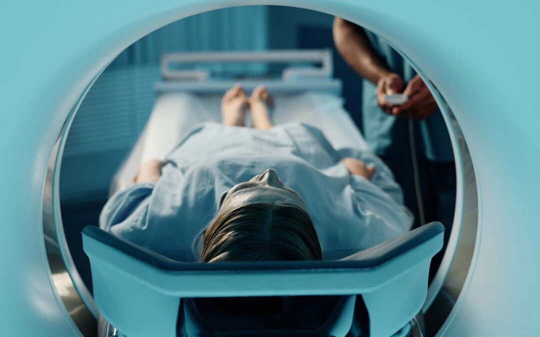
A Computed Tomography (CT) scan is a sophisticated medical imaging technique that combines X-rays and computer technology to produce detailed cross-sectional images of the body. A CT scanner is a large X-ray tube that moves around the body, sending signals to a computer that then creates an image. This technology is useful in a few ways:
- Diagnostic Imaging – A CT scan can find specific changes inside the body that a normal X-ray machine may not detect. They can assess traumatic internal injuries, cancerous tumors, strokes, and more.
- Guidance for Procedures – CT scans can be used to guide minimally invasive procedures, such as biopsies, needle aspirations, or drainage of fluid collections.
- 3D Imaging – The more advanced CT technology creates three-dimensional images useful for surgical planning and evaluating complex anatomical structures.
Computed Tomography is helpful in radiology because the exams only take a few minutes to complete and the images are detailed and accurate. Many medical centers rely on CT machines such as the GE Brightspeed or the Siemens Definition.
Veterinary Radiology Equipment
In veterinary medicine, imaging equipment plays a crucial role in diagnosing and treating a wide range of conditions across different animal species. Veterinary radiology equipment is very similar to the machines used in medical centers for humans. Some equipment is more utilized than others in veterinary settings due to cost, availability, and practicality.
For example, many clinics use X-ray machines to view detailed images of bones, soft tissues, and organs. Portable X-ray machines are becoming increasingly more common as they can be easily maneuvered around veterinary clinics or used in the field for large animals. Ultrasounds are also widely used for imaging of the soft tissue, organs, and pregnancies of animals.
On the other hand, MRI and CT are less common pieces of veterinary radiology equipment due to their cost and size. However, some specialized clinics use this technology for valuable diagnostic information on an animal.
When using radiology equipment in a veterinary setting, clinicians must consider the animal’s size and species, as well as the potential need for restraint. Because veterinary radiology equipment varies in size and capabilities, large animals such as horses may require specialized equipment to complete the scan. Also, these imaging procedures often require animals to be still, which may require animals to go under anesthesia or sedation.
Medical Imaging Systems & Equipment From Imperial Imaging
The Imperial Imaging team is committed to elevating the provider and patient experience with high-quality imaging equipment and exceptional service. Contact us today to discuss imaging solutions that best suit your facility. Our unmatched customer service team will match you with the right machines so you can confidently deliver fast, accurate diagnoses that elevate patient care!

